Most people have never heard of the Lisfranc joint until it fails. Then it becomes the only thing they think about. This injury sits in the middle of the foot at the tarsometatarsal joint complex, and it disrupts everything from walking to driving to standing still. A Lisfranc joint injury is classified as either a sprain, a fracture, or a fracture dislocation. Severity varies widely. Outcomes depend almost entirely on how quickly and accurately the injury is identified and managed. An estimated 1 in 55,000 people per year sustains a Lisfranc injury.
What Does the Lisfranc Joint Actually Do?
The Lisfranc joint is the articulation between the midfoot and forefoot. Five metatarsal bones connect to the cuneiform and cuboid bones through this joint. It is stabilized by a network of ligaments, with the Lisfranc ligament itself connecting the medial cuneiform to the base of the second metatarsal.
This joint transmits enormous forces during walking and running. At push-off, the midfoot bears up to 1.7 times body weight. During sprinting, that force increases significantly. The joint needs to be rigid enough to act as a lever during propulsion while still allowing subtle movement for balance.
When the joint is damaged, the foot loses that rigid lever. Push-off becomes painful or impossible. Arch collapse follows. Every step is compromised.
What Are the Most Common Symptoms?
Symptoms depend on the severity of the injury, but there are clear patterns.
Midfoot pain is the universal complaint. It is worse with weight-bearing and typically eases at rest. Swelling appears across the top of the midfoot, often within minutes of injury. The swelling is usually localized between the first and second metatarsal bases.
Bruising on the bottom of the foot is the most specific clinical sign. It is called plantar ecchymosis. It does not appear in ankle sprains or isolated metatarsal fractures. When a clinician sees it, a Lisfranc injury is the primary concern until proven otherwise.
Inability to bear weight is common in moderate to severe injuries. In mild sprains, some weight-bearing is possible but painful. The ability to walk does not rule out a significant injury. Many patients with torn ligaments still hobble on the injured foot.
How Is the Diagnosis Confirmed?
Diagnosis requires imaging, but the right imaging matters.
Standard non-weight-bearing X-rays miss up to 50% of Lisfranc injuries in some clinical reviews. The joint alignment looks normal when the foot is off-loaded. Weight-bearing X-rays are essential. A gap of 2mm or more between the first and second metatarsals confirms instability.
CT scanning gives detailed information about fracture pattern and bone alignment. It is the preferred imaging for surgical planning.
MRI is most useful in purely ligamentous injuries where no bones are broken. It shows the Lisfranc ligament directly and grades the tear as partial or complete. Complete tears are unstable and require surgical fixation.
Stress X-rays, where the foot is manually stressed while imaging, can also demonstrate instability not visible on standard views. These are used when clinical suspicion is high but routine imaging is inconclusive.
What Are the Treatment Options Based on Severity?
Treatment is not one size fits all. It follows the injury pattern precisely.
Stable sprains with intact ligaments and no displacement are treated conservatively. Six to eight weeks of non-weight-bearing in a cast or boot allows ligament healing. Custom orthotics follow to support the arch during the return to walking.
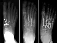
Unstable injuries with displacement require surgery. The debate in orthopedic surgery is between ORIF, where screws and plates hold bones in alignment temporarily, and primary arthrodesis, where the joint is fused permanently.
A 2012 randomized trial in the Journal of Bone and Joint Surgery found that primary fusion produced significantly better outcomes at two years than ORIF for purely ligamentous Lisfranc injuries. Pain scores, function, and return to activity all favored fusion.
For athletes and younger patients, treatment decisions are made with return-to-sport timelines in mind. Fusion eliminates residual instability and often allows a more predictable recovery than ORIF followed by hardware removal.
What Does Long-Term Recovery Really Look Like?
Recovery is measured in months, not weeks. Patients need to hear this early.
At three months, most patients are in footwear with orthotics and doing structured physiotherapy. At six months, functional tasks like climbing stairs and walking longer distances are being worked on. At twelve months, most non-athletes are close to their final functional outcome.
Residual symptoms are common. Arch fatigue, morning stiffness, and sensitivity to prolonged standing affect many patients even at the one-year mark. These symptoms are manageable but rarely disappear entirely in moderate or severe injuries.
Post-traumatic arthritis develops in a significant percentage of cases regardless of treatment method. A 2019 study in Foot and Ankle Surgery reported arthritis rates of 25 to 58% at long-term follow-up across surgical and non-surgical groups.
Ongoing orthotic use plays a real role in managing this. It reduces the load through degenerated joint surfaces and helps maintain arch support when the ligaments can no longer do it alone.